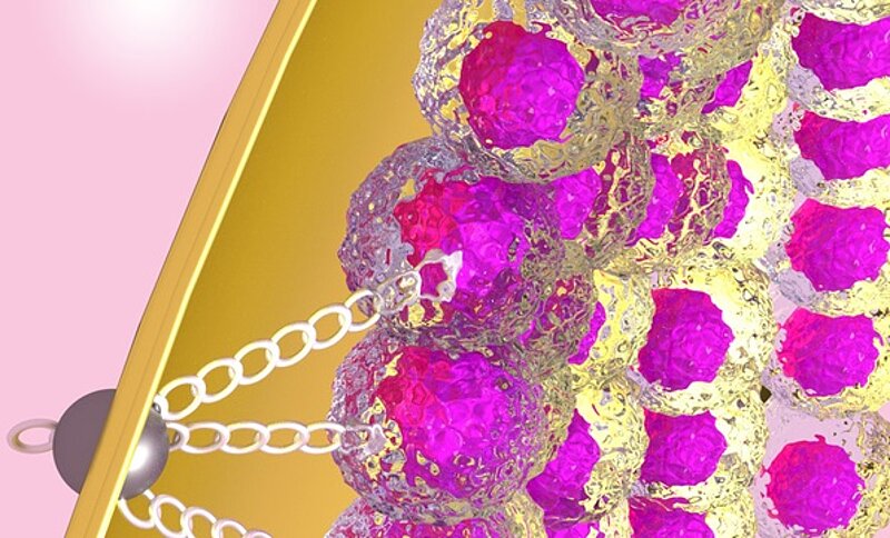
Everyone’s life has its milestones. Lewis Wolpert, a British developmental biologist, once said that it is not birth, marriage or death but gastrulation that is the most important event in life. Gastrulation describes the process during which the single-layered blastula (a hollow sphere of cells) is reorganized into a multilayered structure known as the gastrula. In the process, physical forces reshape the embryonic tissue to form complex body plans of multicellular organisms. In many embryos, the gastrulating tissue is surrounded by a rigid protective shell. So far, scientists did not know whether interactions between the living tissue and the protective shell provide additional forces that affect gastrulation. Studying the red flour beetle, researchers at the Max Planck Institute of Molecular cell Biology and Genetics (MPI-CBG), the Biotechnology Center of the TU Dresden (BIOTEC), and the Cluster of Excellence “Physics of Life” (PoL) recently discovered that the living tissue attaches firmly to the shell that surrounds the embryo. This attachment generates additional external forces that are required for proper gastrulation movements. The study is published in the journal Nature.
Gastrulation is one of the most intensely studied biological processes. During gastrulation, animal embryos undergo tissue flows and folding events that transform a simple monolayer of cells into a complex multi-layered structure called gastrula. This transformation is mainly driven by physical forces generated within the embryonic tissue itself. However, in most species, gastrulation happens within some form of rigid protective shell surrounding the developing embryo. To investigate how the interaction between the living tissue and the inner surface of the protective shell impacts gastrulation, an interdisciplinary team of researchers in Dresden studied this process in the red flour beetle Tribolium castaneum, an emerging model organism exhibiting spectacularly complex tissue movements during gastrulation. They discovered that the tissue attaches to the protective shell in a specific location and that this attachment generates additional external forces that contribute to accurate gastrulation movements.
The research team included biologists from the lab of Pavel Tomancak at the MPI-CBG and physicists from the research group of Stephan Grill who is both a professor at BIOTEC and a director at MPI-CBG. The researchers investigated the tissue movements during the early development of two insects, Tribolium and Drosophila, using Dresden’s signature approach of combining theory and experiment. They first imaged the dynamic development of Tribolium with state-of-the-art light sheet microscopy. Then, they applied a biophysical theory to the measurements and came to a surprising conclusion that an additional force must be involved to explain the gastrulation movements. But what could it be? Stefan Münster, the lead author of the study who was supported by an interdisciplinary postdoctoral fellowship from the Center for Systems Biology Dresden (CSBD), explains the discovery: “We found through embryological experiments that the unknown force originated from the attachment of the gastrulating tissue to a specific location of the protective shell. We then figured out the molecular components required for this attachment and showed that they also play a role in the gastrulation movements of the fruit fly Drosophila.”
These findings uncovered a thus far overlooked mechanism contributing to the physical reshaping of tissues during animal development. Bridging physics and biology, Stefan Münster worked with physicist Alexander Mietke and biologist Akanksha Jain on both the theoretical modelling and experimental validation of the proposed attachment mechanism. “It was an exciting journey from physics to biology and back”, says Stefan Münster. “We were the first to use the red flour beetle for biophysical investigations. By looking at this beetle, we learned something new about the tissue flow during gastrulation – the anchoring of the tissue to the shell. This interaction has so far been missed when looking only at Drosophila”, summarizes Stefan Münster the findings. Stephan Grill adds: “Dresden is an ideal place to pull of such a study. We have biologists studying diverse model species at the MPI-CBG and theorists capable of modelling the physics of biological processes at the CSBD. In fact, we are just starting a new institute at TU Dresden, the Cluster of Excellence “Physics of Life”, focusing on exactly these types of interdisciplinary topics.”
Besides physics, these results also open new perspectives on how gastrulation may have evolved between species. “Despite being a signature event of early development that sets the tone for the rest of the animal’s life, gastrulation is very flexible and even related species show widely different gastrulation folds and movements” says Pavel Tomancak. “How and why gastrulation evolved so rapidly is a long-standing mystery in evolutionary biology.” The work of the Dresden team points towards localized interaction of the living tissue with its protective environment as one of the mechanisms contributing to the diverse ways, an animal can develop.
Original publication:
Stefan Münster, Akanksha Jain, Alexander Mietke, Anastasios Pavlopoulos, Stephan W. Grill & Pavel Tomancak: “Attachment of the blastoderm to the vitelline envelope affects gastrulation of insects” Nature, 27. March, 2019. doi: 10.1038/s41586-019-1044-3
