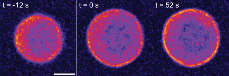
The discovery by researchers at the Cluster of Excellence Physics of Life in Dresden (PoL) sheds light on a crucial “safety belt” mechanism that protects the structural integrity of mammalian cells, allowing them to withstand mechanical stressors. The study, led by Prof. Dr. Elisabeth Fischer-Friedrich in collaboration with Prof. Dr. Michael Schlierf’s research group at the Center for Molecular Bioengineering (B CUBE), both located in Dresden, Germany, reveals the remarkable adaptability and resilience of the actin cortex, a shield-like structure on the outer layer of mammalian cells.
The actin cortex, composed of micron-sized filaments formed by the actin protein and interconnected by molecular cross-linkers, acts as a dynamic shield for a cell, regulating cellular shape through various stages of its lifecycle. This unique structure must strike a delicate balance, being both flexible enough to enable changes in its form and composition and robust enough to prevent cell rupture. Until now, the mechanisms governing the actin cortex's response to tension remained a mystery.
When subjected to stress, the actin cortex enhances its structural integrity by generating additional actin filaments and cross-link components. In a remarkable analogy, researchers likened this mechanism to a “safety belt” for cells, in which the actin cortex strengthens itself in response to mechanical tension similar to the safety belt in a car. Valentin Ruffine, the first author of the research paper, highlights the significance of this discovery, stating, “We see a mechanosensitive response both at the level of the cross-linker proteins and in the actin polymerization.”
Prof. Dr. Elisabeth Fischer-Friedrich, head of the research group, emphasizes the significance of their findings, saying, “The increase in density of cross-links and actin filaments over time due to mechanical forces in the cortex was a key revelation, providing a deeper understanding of how cells adapt their material properties in response to mechanical challenges such as rapid deformation.”
This pioneering research was conducted by subjecting living cells to controlled forces using atomic force microscopy and employing high-resolution imaging techniques, complemented by mathematical modeling.
The study's findings represent a significant leap forward in our understanding of cellular mechanics and offer hope for innovative approaches to addressing diseases linked to cellular shape regulation, cell migration, and tissue organization. This groundbreaking research exemplifies the ongoing commitment of scientists at PoL and B CUBE to unravel the mysteries of life at the cellular level.
