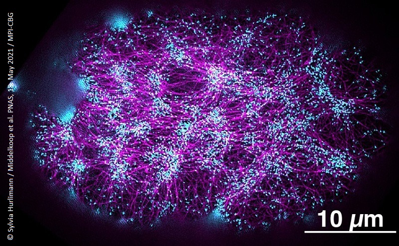
Our internal organs are arranged left-right asymmetrically, which is crucial for an embryo to grow properly and to define our body plan. In a collaboration between Dresden, Boston and California, researchers now identified proteins that create a molecular torque - a rotary force - on the cellular level that plays an important role to create this left-right asymmetry during development, ultimately shaping the body plan. Stephan Grill summarizes: “These findings are another example of bridging physics and biology that has long been done in an ideal way here in Dresden”.
See also: Press release "Rotating forces get embryos in shape" (on website of MPI-CBG)
Original publication: CYK-1/Formin activation in cortical RhoA signaling centers promotes organismal left-right symmetry breaking. PNAS, 18. May 2021, Doi: 10.1073/pnas.2021814118
