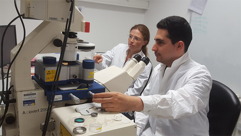
Scientists led by Dr. Elisabeth Fischer-Friedrich, group leader at the Cluster of Excellence Physics of Life (PoL) of TU Dresden, discover how epithelial cells change their mechanical properties upon the so-called epithelial-mesenchymal transition (EMT). This cellular transition is known as a hallmark of cancer progression and malignancy. In particular, the researchers looked at cell lines derived from breast, lung and prostate tissue - tissues that exhibit cancerogenic transformations more frequently than others. Kamran Hosseini from the Fischer-Friedrich lab comments: “Our findings open up new ways to identify EMT in cancer cells and to reconsider its role in cancer progression such as tumor growth and metastasis.”
Epithelial cells line the surface of organs in the body of humans and animals. Some epithelial cells undergo transformations and constitute a type of cancer called carcinoma, which make up the majority (~85%) of malignant tumors in humans. A cellular transformation named epithelial-mesenchymal transition (EMT) has been characterized as a hallmark of cancer progression and metastasis. As part of this transition, epithelial cells reorganize their internal structure and weaken their attachment to neighboring cells. “EMT therefore promotes cell migration and cancer cell invasiveness during metastasis,” says Dr. Kamran Hosseini, first-author of the study. “We were curious to which extent EMT influences the mechanical properties of the cell. Therefore, we characterized the cells using an atomic force microscope,” explains Dr. Kamran Hosseini. They used this state-of-the-art setup to measure physical properties such as cell stiffness and cell surface tension before and after EMT. The authors observed that EMT influenced the cancer cells in two contrasting ways, depending on whether the cell was in the process of dividing: they discovered a softening in interphase but a stiffening in mitosis upon EMT.
“We plan to further characterize the molecular changes of the cytoskeleton that lead to the mechanical phenotype we observed. These results would not only serve as important insight on cell-biological regulation, but could also aid future research to identify new targets for cancer therapeutics,” says Dr. Elisabeth Fischer-Friedrich.
Original publication:
Kamran Hosseini, Annika Frenzel, Elisabeth Fischer-Friedrich: "EMT changes actin cortex rheology in a cell-cycle-dependent manner", Biophysical Journal, 17 August 2021, doi: https://doi.org/10.1016/j.bpj.2021.05.006
Further information:
Dr. Elisabeth Fischer-Friedrich
Group leader for Mechanics of Active Biomaterials
DFG Cluster of Excellence Physics of Life (PoL), TU Dresden
Phone: +49 351 463-40325
Email: elisabeth.fischer-friedrich@tu-dresden.de
Media contact PoL:
Bianka Claus
Public relations
DFG Cluster of Excellence Physics of Life (PoL), TU Dresden
Phone: +49 351 463-40133
Email: bianka.claus@tu-dresden.de
